 NCS
and Native Patterson MapsNCS
and Native Patterson Maps
NCS
and Native Patterson MapsNCS
and Native Patterson MapsSearch for non-crystallographic symmetry (NCS) with Patterson self-rotation maps does not reveal any new peaks in cases where the NCS axis is parallel (or nearly so) to a crystallographic axis of same or higher symmetry in the respective spacegroup. Occurrence of NCS parallel to crystallographic axes is not uncommon. Such a NCS axis can be found by inspecting native Patterson maps. All you need to do is create a Patterson map from the data and a large peak will occur. The position of the axis can be calculated from the Patterson peak position. I found no description in a basic textbook, but see Meth. Enz. 277, p26, and the reference therein
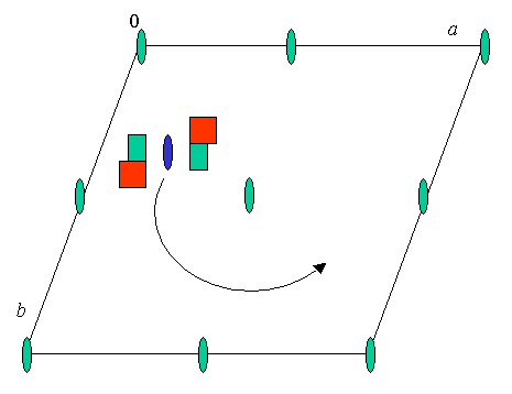
Let us look at a monoclinic cell, P2 (No.3) for example, with two molecules (red-green square protein, RGSP-1) in the asymmetric unit related by a twofold NCS axis (blue symbol) parallel to the twofold crystallographic axis (green). Standard setting, unique axis b. The origin could be at any of the 2-fold axes.
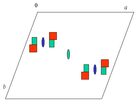
Applying any of the equivalent 2-fold operations creates a second copy of the 2 molecules (RGSP-1 dimer) in the unit cell :
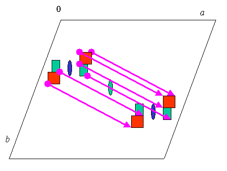
As you may have seen already, there is now a set of translational vectors relating each atom in the 2 copies of the molecule (I have shown only a few vectors) :
The large number of same (or very similar, in non-perfect NCS) distance vectors of course gives rise to a whopping peak in the native Patterson map :
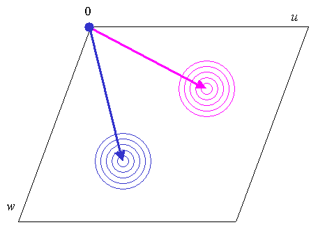
The blue peak is symmetry related to the purple one, and directly gives the position of the NCS axis from the general Patterson peak position at u,v,w = 2x, 0, 2z. From the way I drew the vectors between the molecules in the unit cell the purple peak actually yields the origin shifted solution: translate origin to 1/2,0,1/2 and then add the x and z derived from the purple vector (the origin can be chosen at any of the 2-fold axes in P2).
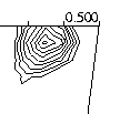
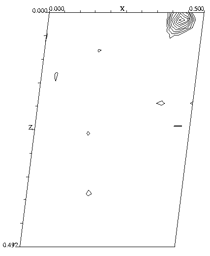
Click here for a real life example (native Patterson NCS peak in P21 , 2E8 Fab antibody fragment) 1
QUIZ
Where would the NCS peak be in space group P21 ?
![]() Back to X-ray Tutorial Index
Back to X-ray Tutorial Index
This World Wide Web site conceived and maintained
by Bernhard Rupp.
Last revised
Dezember 27, 2009 01:40